
Harmful effects of ammonia on birds
Ammonia gas in poultry houses seriously affects the health condition of the birds. Good litter management and ventilation will minimise the level of ammonia, improve productivity, reduce the likelihood of respiratory diseases, improve the birds’ welfare and provide a pleasant, safe environment for workers.
By Dr. Tahseen Aziz, Rollins Animal Disease Diagnostic Laboratory, North Carolina Department of Agriculture and Consumer Services, and Dr. H. John Barnes, College of Veterinary Medicine, NC State University, Raleigh, NC, USA
Ammonia is a gas present in the atmosphere of every poultry house. It results from the chemical decomposition of uric acid in droppings by certain bacteria in the litter. It is particularly high in houses where the same litter is used for successive flocks. The main factors affecting atmospheric ammonia concentration in poultry houses are litter conditions and air movement (ventilation). Moisture content, pH and the temperature of the litter, influence the degradation of uric acid by bacteria. Poor ventilation, loose droppings and faulty, over filled or low positioned drinkers, are common causes of wet litter in poultry houses.
General effect of ammonia gas
Ammonia gas has a characteristic pungent odour. At high concentrations it is irritating to mucous membranes of the respiratory tract and the conjunctivae and corneas of the eyes. Damage to the mucous membranes of the respiratory system increases the susceptibility of birds to bacterial respiratory infection, especially E. coli infection. High levels also have a negative impact on overall livability, weight gain, feed conversion, condemnation rate at processing and the immune system of the birds. Experimentally, broiler chickens kept in an environment with ammonia concentrations of 50 ppm and 75 ppm were shown to have reductions in body weight of 17% and 20%, respectively, at 7 weeks of age compared to broiler chickens kept in an environment with near 0 ammonia concentration. In the US, maximum levels of ammonia in poultry houses have been set at 25 ppm by the National Institute of Occupational Safety and Health (NIOSH) and 50 ppm by the Occupational Safety and Health Administration (OSHA). These levels have been established based on human safety and represent the limits for 8 hours of exposure. OSHA considers 50 ppm to be the lowest level to cause irritation to the eyes, nose and throat of the most sensitive individuals. People can generally smell ammonia at concentrations between 20 and 30 ppm.
Damage to the respiratory system
The effect of ammonia gas on the mucosal surface of the trachea ranges from paralysis of cilia, to deciliation (loss of cilia) of epithelial cells, to injury (necrosis) of the mucosal epithelium itself. Attenuation of the mucosal epithelium, with loss of cilia and increased numbers of goblet cells, are common lesions of aerial ammonia toxicity seen in the tracheas of affected birds. The type and degree of damage depends on the concentration of ammonia in the air and the length of time of exposure. Cilia are tiny, hair-like projections on the surface of respiratory epithelial cells that line the trachea. They form part of the “mucociliary apparatus” (also called mucociliary blanket or mucociliary escalator), which consists of the cilia and secretion of mucus. The mucociliary apparatus is responsible for entrapping and cleaning particles inhaled from the environment. Dust particles have large numbers of bacteria attached to their surface. Poultry house dust has been shown to be a reservoir for E. coli in particular. Inhaled particles are entrapped in mucus secreted by mucous glands and goblet cells. Propulsive actions of cilia continuously move the mucus with entrapped particles up the trachea toward the pharynx, thus preventing the particles from reaching the lower respiratory tract (lungs and air sacs). Clearance of particles from the respiratory tract of birds is approximately 30 times faster than that of mammals. When cilia become paralyzed or are lost due to high ammonia levels in the poultry house, mucus on the mucosal surface of the trachea cannot be cleared, and thus entrapped bacteria on dust particles may reach the lungs and air sacs and cause infection. Devitalisation of the tracheal mucosa may explain in part the high incidence of airsacculitis, pneumonia and septicemia caused by E. coli in poultry flocks that have been exposed to high atmospheric ammonia levels. Prolonged exposure to atmospheric ammonia may also incite proliferation of the epithelial cells that line the atrial spaces in the lung. The proliferative lesion causes thickening of parabronchial walls. In severe cases, total obliteration of atrial spaces, with subsequent reduction in pulmonary gas exchange can occur.
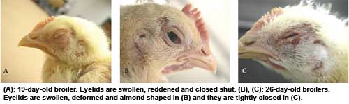
Damage to the eyes
Atmospheric ammonia at high concentrations causes conjunctivitis (inflammation of conjunctivae) and damages the cornea of the eyes. As in the respiratory system, the severity of damage depends on the concentration of ammonia in the environment and duration of exposure. Swelling and reddening of the eyelids, reddening of the conjunctive and nictitating membrane (third eyelid), and partial or complete closure of the eyes are common clinical signs. In severe cases, the eyelids are often closed shut. Eyes can become almond shaped after longterm exposure to high ammonia levels because of scarring and retraction of the eyelids. Conjunctivitis caused by ammonia also increases the risk and severity of swollen head syndrome in respiratory viral infections. Histopathologic lesions in conjunctivae include hyperemia (congestion of blood vessels with blood), variable degrees of disorganisation and hyperplasia of conjunctival epithelium, and mild to moderate infiltration of heterophils. In the eyelids, there may be edema, with subepithelial infiltration of heterophils.
Lesions in the cornea
The term “ammonia burn” is used by some to refer to ammonia-induced corneal erosion, but this term is inaccurate, as ammonia does not directly injure the corneal epithelium. Damage to the basement membrane on which the epithelium rests is responsible for detachment of the epithelial layer, which is the characteristic eye lesion that results from excess ammonia exposure. The lesion is an almost circular, grey-white, opaque, rough-looking area in the centre of the cornea. Close examination of the eye using oblique light may be necessary to see early lesions that are slightly depressed with an irregular margin. The periphery of the cornea is unaffected, presumably because it is partially covered by the eyelid and receives less exposure than the central cornea. Both eyes are similarly affected, which helps to differentiate ammonia toxicity from other eye diseases. These birds are partially to completely blind. Histologically, the corneal lesion is an erosion in which there is separation and loss of the corneal epithelium from the underlying basement membrane in the central cornea. Along the margins of the erosion, the corneal epithelium is lifted from the basement membrane. In some cases, which probably represent early lesions, the corneal epithelium is still present, but it is separated from the basement membrane. The denuded basement membrane is thickened and basophilic because of the deposition of calcium salts. The resulting band of mineralized basement membrane is known as “calcifyc band keratopathy”. It is characteristic of ammonia toxicity in poultry. How ammonia induces band keratopathy, which subsequently results in detachment of the epithelium from the basement membrane, is unknown. In people and some other animals, calcific band keratopathy occurs as a complication of uveitis (inflammation of the middle tunic of the eye composed of the choroid, iris and ciliary body).
The cornea usually remains intact and does not perforate. In chronic lesions, there is fibroplasia in the corneal stroma under and around the eroded area, and blood vessels extend into the cornea (normally there are no blood vessels in the cornea). Regenerating epithelium usually covers the fibroplastic area indicative of a healing process. Another lesion commonly seen in affected eyes is varying degrees of iritis (inflammation of the iris) characterized by infiltration by lymphocytes, plasma cells, and some granulocytic leukocytes. Mild, mixed inflammatory cell infiltrate may also present in the ciliary body. Band keratopathy and corneal erosion may be secondary to these inflammatory changes in the uvea.
Join 31,000+ subscribers
Subscribe to our newsletter to stay updated about all the need-to-know content in the poultry sector, three times a week.


 Beheer
 WP Admin
  Bewerk bericht