
Infectious Laryngotracheitis (ILT) targets broilers
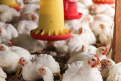
Infectious Laryngotracheitis is a viral disease most commonly found in chickens, but is also seen in other fowl. In broilers, the vast majority of outbreaks occur in flocks over 45 days of age, but the disease has been diagnosed in flocks of a younger age.
By Dr Tahseen Aziz, Avian pathologist, Rollins Animal Disease Diagnostic Laboratory, North Carolina Department of Agriculture and Consumer Service, Raleigh, NC, USA
Infectious laryngotracheitis (ILT) is a viral respiratory disease caused by a herpes virus. Natural infections with ILT virus occur mainly in chickens, and both young and adult chickens are susceptible to infection. In broilers, for uncertain reasons, the vast majority of outbreaks occur in flocks over 45 days of age. However, ILT has been diagnosed in broiler chickens as young as three weeks. Natural ILT virus infections rarely affect pheasants and peafowl, but have recently been reported for the first time in turkeys.
Clinical picture
Typically, there is a sudden increase in daily mortality in a single house on a farm. Often, birds in one area of the house show clinical signs and die, and the disease spreads relatively slowly through the other birds in the house. Daily mortality progressively increases and can affect hundreds of birds each day. Severity of clinical signs and mortality rates vary widely among outbreaks depending on the immune status of the flock, initial exposure dose, age of the birds, virulence of the viral strain, co-infections with other respiratory pathogens, and environmental factors that adversely affect the respiratory system. Outbreaks are generally more severe in older rather than younger broiler flocks.
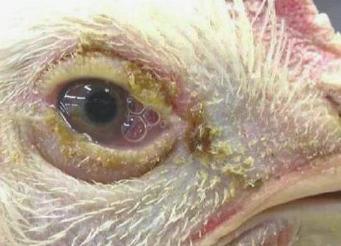
Infected birds are lethargic and have swollen eyelids, reddened conjunctivae, and watery eyes. In severe cases, eyelids may be adhered with dry, crusty, ocular discharge sticking to the eyelids and skin around the eye. Severely infected birds exhibit mouth breathing, coughing, and gargling respiratory noises. Some birds stretch out their neck while trying to breath. Coughing up bloody mucus is a characteristic clinical sign that occurs in birds with badly damaged tracheas; bloody mucus may be found on and around the beak, and in the oral cavity. Sp ots of dried bloody exudate may be found in the bird’s environment on sidewalls and equipment when flocks are severely affected.
Lesions in the lungs, if present, are notspread but rather multifocal or regionally extensive. The normal histologic architecture in affected areas is severely distorted. Focal or diffuse areas of the mucosal epithelium of primary and secondary bronchi a re either lost or disorganised. Walls of affected bronchi are usually infiltrated with large numbers of inflammatory
mononuclear cells, and exudate c omposed of fibrin, heterophils, and mononuclear cells in different proportions is usually present in the bronchial lumen. Parabronchi, atria, and capillary beds around affected bronchi typically show marked proliferation, lymphohistiocytic inflammatory response, fibrin admixed with heterophils, and obliteration o f air spaces. Syncytial cells with intranuclear inclusions may be seen in the lesions, but large syncytial cells without nuclear inclusions are also commonly encountered. Het erophilic, caseating granulomas may be present.
Join 31,000+ subscribers
Subscribe to our newsletter to stay updated about all the need-to-know content in the poultry sector, three times a week.


 Beheer
 WP Admin
  Bewerk bericht